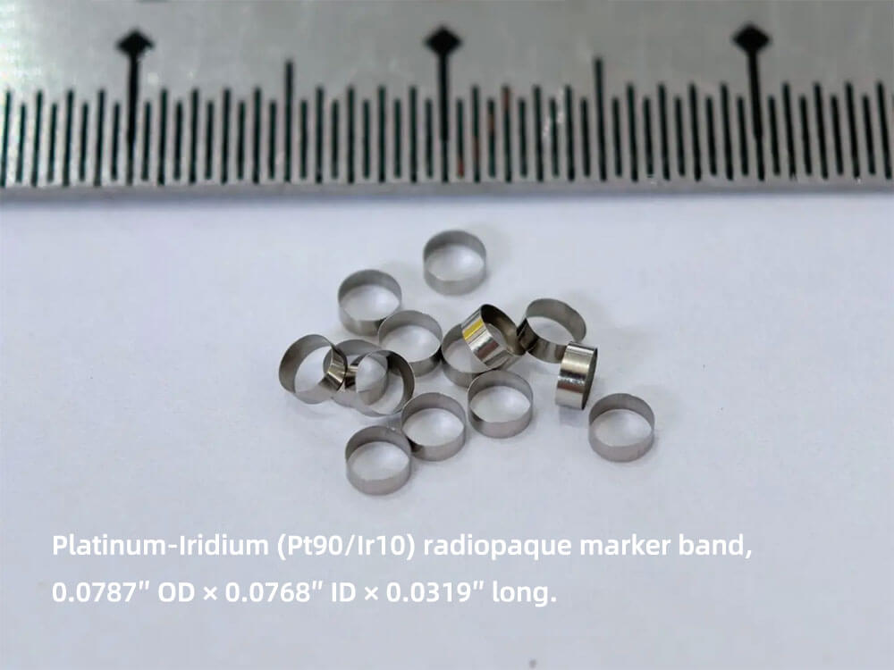

Think of a marker band as a tiny “headlight” on a catheter. It’s nothing more than a miniature ring—usually smaller than a grain of rice—crimped onto the shaft. The trick? The ring is made from heavy metals such as platinum or a platinum-iridium alloy. Those metals are much denser than blood, tissue, or even bone, so when the X-ray beam hits them they soak up more radiation and cast a bright white shadow on the fluoroscopy screen. That white dot is the doctor’s GPS; it shows exactly where the tip of the device is in real time.
| What’s Happening | How We Do It | What the Doctor Sees |
|---|---|---|
| X-ray absorption | Slip a Pt/Ir ring over the catheter | A crisp white line against the gray anatomy |
| Exact positioning | Place the band at a known distance from the tip | Live “you-are-here” guidance while the device moves |
Quick Walk-Through
- Patient rolls into the cath-lab.
- Surgeon advances the catheter.
- Fluoro X-ray switches on—bam, the ring glows white.
- Doctor steers until the white dot reaches the target (lesion, vessel, stone, etc.).
- Therapy happens—balloon inflated, stent deployed, cement injected—done.
Because platinum doesn’t rust, react, or leach, the ring can stay in the body forever without causing trouble. Outside the heart, the same idea pops up in spine-balloons (vertebral augmentation) and kidney-stone baskets (multiple bands = 3-D map). Same principle, different body part.
Bottom line: heavy metal + X-ray = instant roadmap. No batteries, no wires—just physics doing the navigation.
Process Explanation:
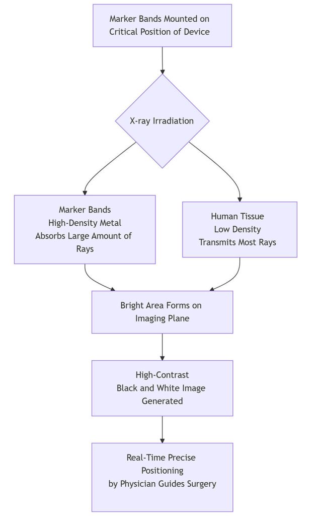
- Device Preparation (Marker Bands Mounted on Critical Position of Device): The marker bands are fixed at key positions on interventional medical devices, such as the distal end of a catheter, balloon, or stent.
- X-ray Irradiation: During surgery, an X-ray source penetrates the target area of the human body.
- Differential Absorption (Key Step):
- Marker Bands Absorb Rays: The high-density metals (such as platinum-iridium alloy) used in the marker bands have a strong ability to absorb X-rays.
- Human Tissue Transmits Rays: Human soft tissues and blood have lower density, allowing most X-rays to pass through.
- Image Contrast Formation: The differential absorption and transmission of X-rays result in dark areas (where the marker bands are located) and bright areas (surrounding tissues) on the receiver (e.g., a fluorescent screen or digital detector).
- Image Generation: The contrast between light and dark areas forms a high-contrast black-and-white image.
- Surgical Guidance: Physicians observe the clear position of the marker bands in the real-time image to accurately guide the instrument to the lesion and perform the operation.
💡 Key Technical Details
- Core Principle: Creates image contrast by leveraging differences in material density and their resulting differential absorption of X-rays.
- Material Selection: Commonly used materials include platinum-iridium (Pt-Ir) alloy, tantalum (Ta), and gold (Au). These metals are chosen for their high density (providing good radiopacity), excellent biocompatibility, and chemical stability.
- Manufacturing Precision: The manufacturing of marker bands is an art of micron-level precision. Advanced processes like “vacuum melting, laser cutting, and ultrasonic polishing” are required to achieve the necessary high precision and surface quality.